Merkel Cell Carcinoma Immunohistochemistry
The number of granules in case 2 however was. Register to See Applicable Discounts.
Pathology Outlines Merkel Cell Carcinoma
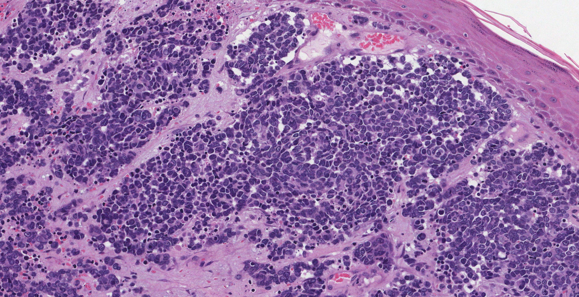
Merkel cell carcinoma is a rare cutaneous neuroendocrine tumor that occurs predominantly in the head and neck region of older patients.
Merkel cell carcinoma immunohistochemistry. 1Department of Laboratory Medicine and Pathology Mayo Clinic Rochester MN 55905 USA. In terms of the remaining antigens there were no differences of significance between the two neoplasms. Immunohistochemistry is often used to confirm Merkel cell carcinoma MCC.
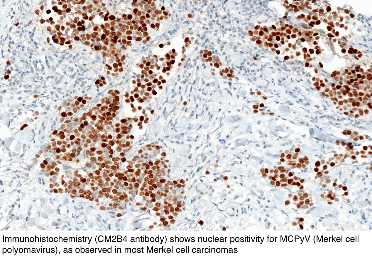
Agnosis frequently requires support by immunohistochemistry. Merkel cell carcinoma a rare cutaneous neuroendocrine tumor of the skin can be categorized into two groups according to Merkel cell polyomavirus MCV presence. Merkel cell carcinoma with heterologous rhabdomyoblastic differentiation.
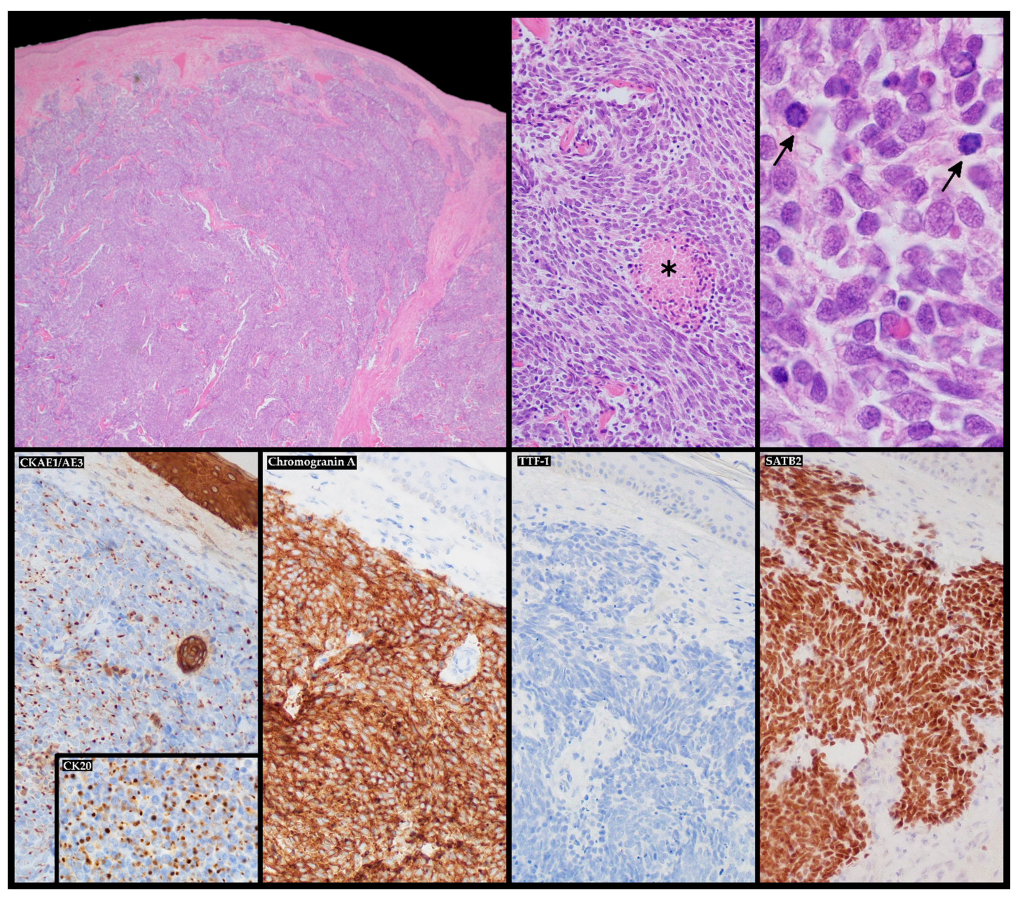
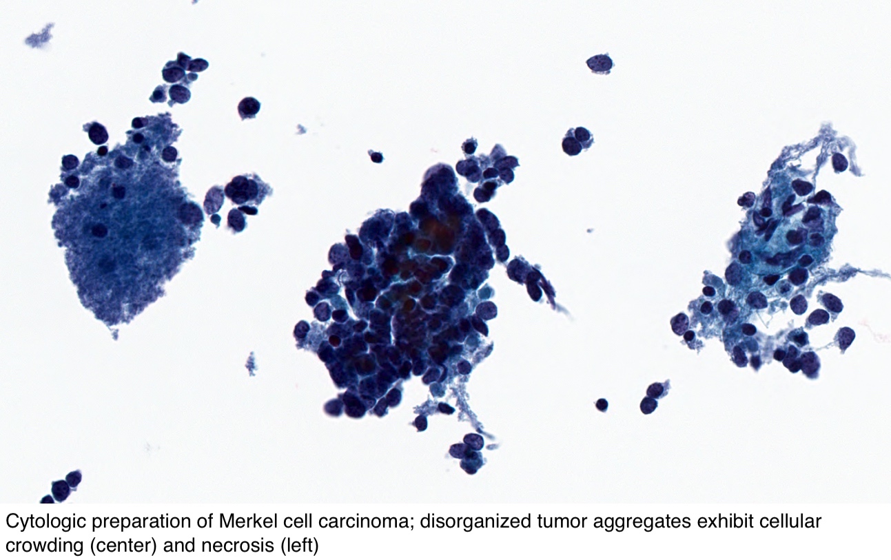
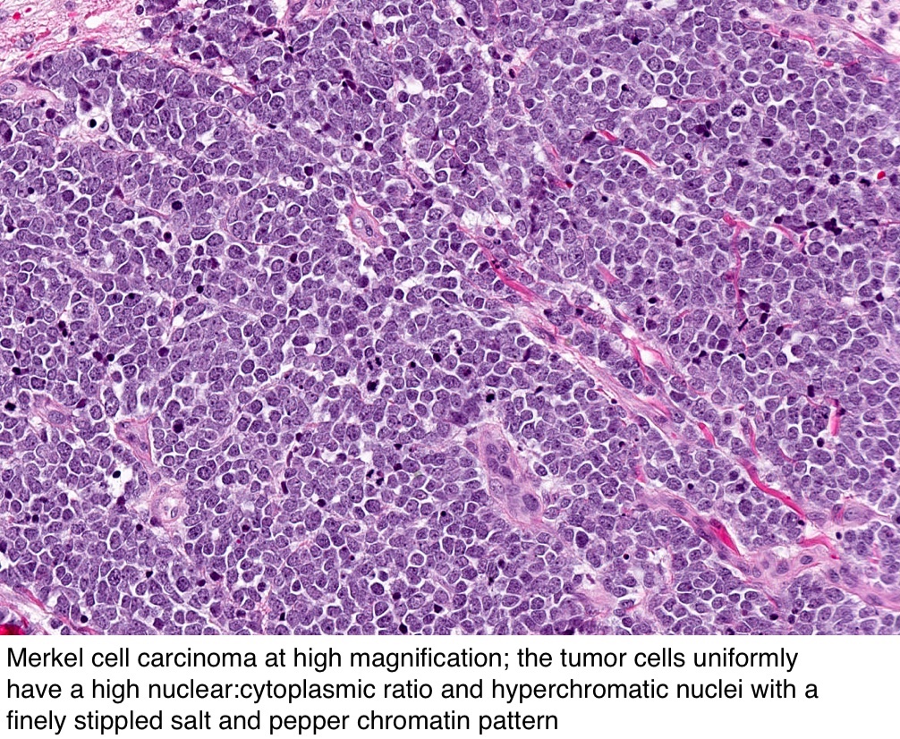
Histologically tumor cells which extended from the dermis into the subcutis showed anastomosing bands with partial trabecular pattern. Reactivity with anti-CK 20 was demonstrated in 23 of 26 Merkel cell carcinomas 88. Merkel cell carcinoma MCC is a cutaneous neoplasm histopathologically difficult to differentiate from other small blue cell neoplasms.
Ultraviolet radiation immunosuppression and the Merkel cell polyomavirus MCPyV are thought to be causative factorsThe cell of origin remains debatable but the immunohistochemical profile and morphology resemble native Merkel cells in the skin. Merkel cell carcinoma is an aggressive tumour that usually arises on chronically sun exposed skin of the elderly. Two cases of Merkel cell carcinoma MCC were examined by electron microscopy and immunohistochemistry.
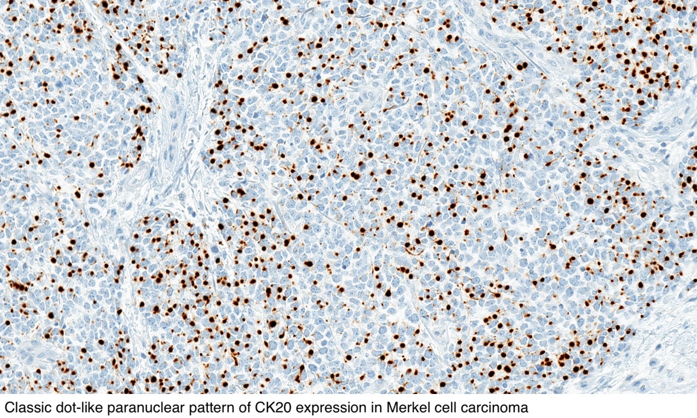
Cytokeratin 20 CK20 positivity is currently used to distinguish. The role of immunohistochemistry for Merkel cell polyomavirus large T-antigen in confirmation. Results Ten of 11 MCCs stained with the antibody to CK20.
Immunohistochemistry for Merkel cell polyomavirus is highly specific but not sensitive for the diagnosis of Merkel cell carcinoma in the Australian population Author links open overlay panel Julie Y. All three CK 20negative tumors showed punctate staining. Negativity for MCV suggests that the virus is not implicated in the development of this subtype of MCC.
Merkel cell carcinoma of the skin primary neuroendocrine cell carcinoma of the skin Clinical features Typically a rapidly growing lesion on sun-damaged skin of the head and neck or extremities of the elderly 10Clinical diagnoses are commonly erroneous. Gill FRCPA a e. Mainos A New Era for Immunohistochemistry.
Merkel cell tumors stain positively for NSE as would any APUD cell. TAp63 tumor suppressorlike properties and Np63 oncogenic properties. MCV-negative tumors are more aggressive and frequently associated with gene mutations.
P63 immunohistochemistry marks the 2 main isoforms of this transcriptional protein. An 88-year-old white man had an erythematous umbilicated tumor on his lower lip which on histopathologic examination showed solid sheets of infiltrating basaloid round cells with a high mitotic index. Methods Eleven cases of MCC and 10 of lung SCC were stained for CK20 and TTF-1.
Recently cytogenetic analysis has emerged as a potential tool in the diagnosis of solid neoplasms including MCC. These findings suggest that a set of three immunohistochemical stains including CK20 NF and TTF-1 is useful in affording a distinction between Merkel cell carcinoma and small cell lung carcinoma. The authors investigated cytokeratins CKs 56 7 17 and 20 staining in paraffin sections of 26 Merkel cell carcinomas to expand the knowledge of the CK staining profile of this entity.
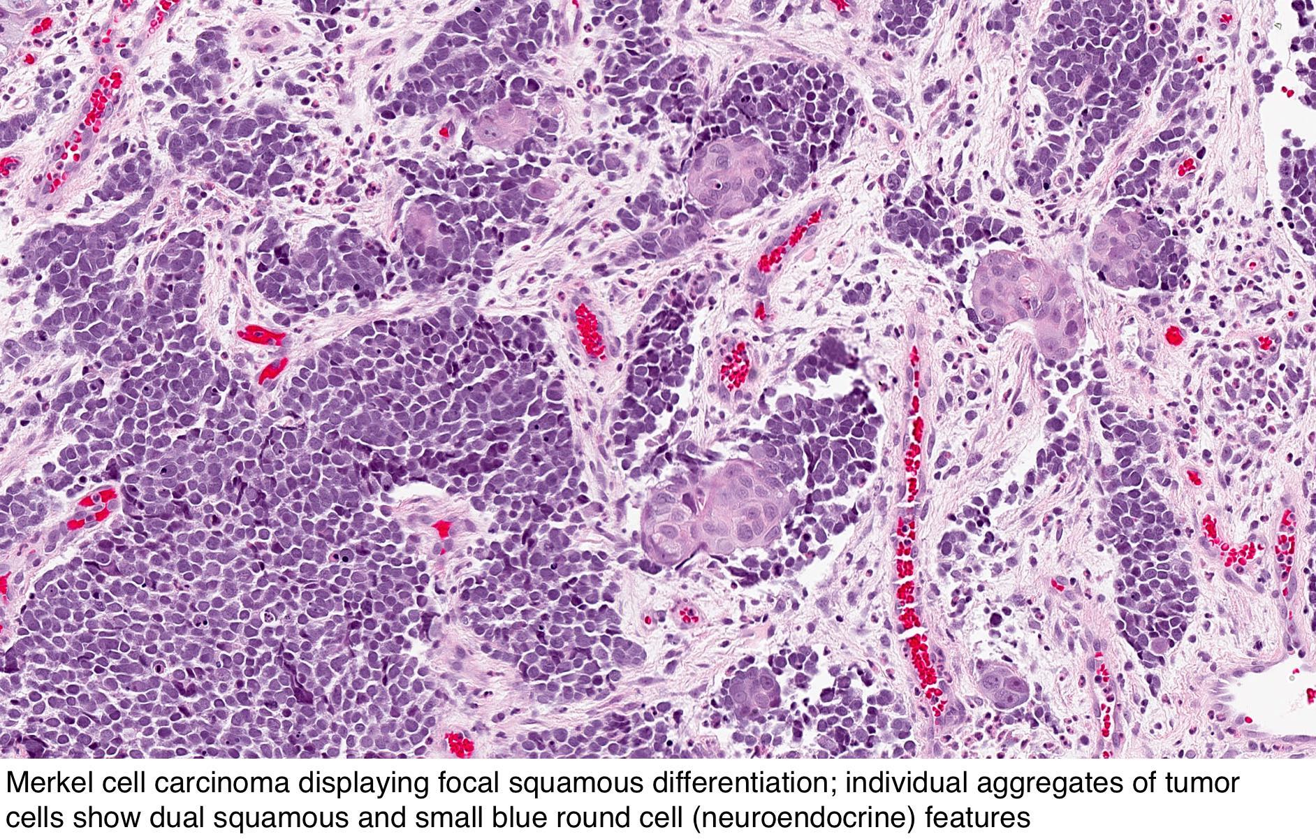
Register to See Applicable Discounts. Aim To investigate whether immunohistochemical staining for cytokeratin 20 CK20 and thyroid transcription factor 1 TTF-1 is useful in distinguishing Merkel cell carcinomas MCCs from metastatic small cell carcinomas SCCs. Merkel cell carcinoma with divergent differentiation is a highly aggressive tumour that might be difficult to recognize owing to its wide histological variability.
The literature suggests that p63 expression in Merkel cell carcinoma MCC is associated with a poor prognosis. Merkel cell carcinoma is a highly aggressive primary cutaneous neuroendocrine carcinoma primarily affecting elderly and immunosuppressed individuals. Diagnosis requires microscopic evaluation as the clinical appearance is nonspecific and can mimic a variety of benign and malignant skin lesions.
Mainos A New Era for Immunohistochemistry. Adhikari LA1 McCalmont TH Folpe AL. Paik MBBS a Geoffrey Hall FRCPA b Adele Clarkson BSc a Lianne Lee MBBS c Christopher Toon MBBS d Andrew Colebatch MBBS a Angela Chou FRCPA c Anthony J.
Immunohistochemical and ultrastructural analyses are usually helpful in differentiating these neoplasms. The ultrastructural study showed tumor cells in case 1 with numerous neurosecretory granules. Merkel cell carcinoma is a rare neuroendocrine carcinoma of the skin mostly induced by Merkel cell polyomavirus integration.
Pathology Outlines Merkel Cell Carcinoma
Pathology Outlines Merkel Cell Carcinoma
Merkel Cell Carcinoma Mypathologyreport Ca
Pathology Outlines Merkel Cell Carcinoma
Pathology Outlines Merkel Cell Carcinoma
Ijms Free Full Text Merkel Cell Carcinoma From Molecular Pathology To Novel Therapies

Immunohistochemistry Of Merkel Cell Carcinoma Metastases 400 Original Download Scientific Diagram

Pathology Outlines Merkel Cell Carcinoma

Researchers Unlock Keys To Staging And Risk Stratification Of Merkel Cell Carcinoma Consult Qd

Tumor In Right Thigh Histology Reveals Merkel Cells Carcinoma Download Scientific Diagram
![]()
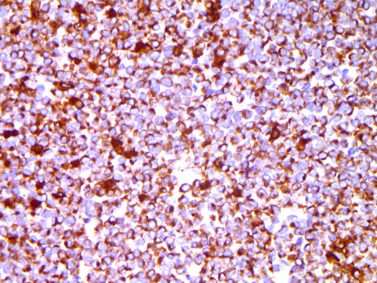
Ck20 Staining Showing The Characteristic Perinuclear Dot Like Positivity Download Scientific Diagram
Pathology Outlines Merkel Cell Carcinoma

Clinical Features And Outcomes Of Merkel Cell Carcinoma In 20 Cats Sumi 2018 Veterinary And Comparative Oncology Wiley Online Library

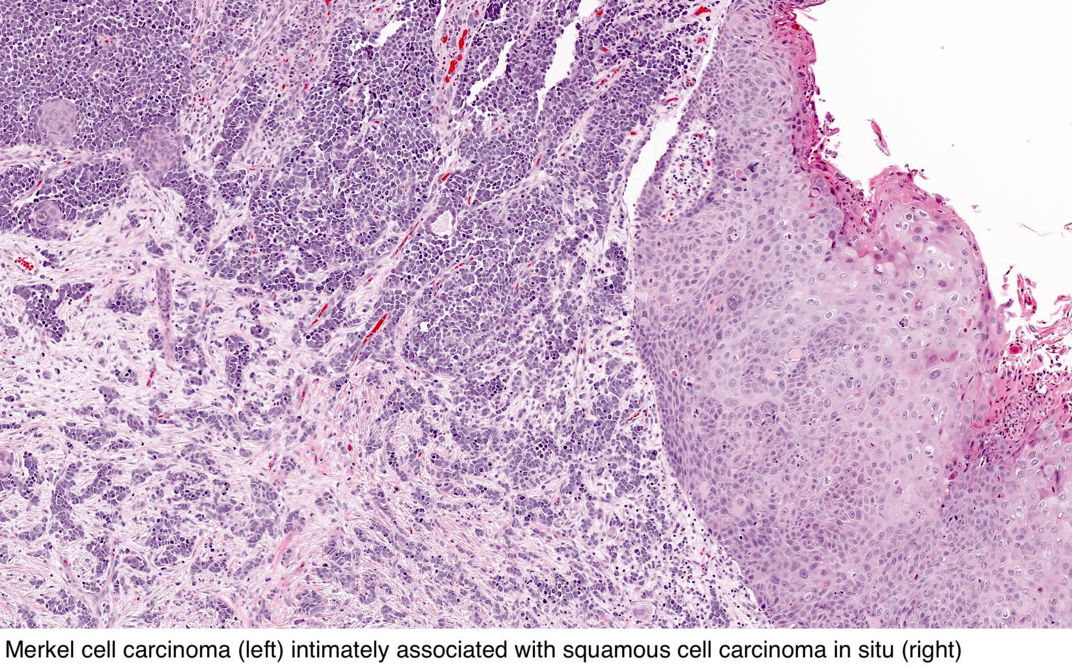
Intraepidermal Merkel Cell Carcinoma With Pagetoid Bowen S Disease Miraflor 2016 Journal Of Cutaneous Pathology Wiley Online Library

An Update On Diagnostic Features Of Merkel Cell Carcinoma Diagnostic Histopathology
Brendan Dickson Md On Twitter Merkel Cell Carcinoma Ihc Ck20 Chromogranin Nb Sheets And Nodules Of Basophilic Cells Granular Chromatin

The Origins Of Various Types Of Skin Cancer 1 Merkel Cell Merkel Cell Download Scientific Diagram

Merkel Cell Carcinoma Can Be Distinguished From Metastatic Small Cell Carcinoma Using Antibodies To Cytokeratin 20 And Thyroid Transcription Factor 1 Journal Of Clinical Pathology

Immunohistochemistry Of Merkel Cell Carcinoma Metastases 400 Original Download Scientific Diagram